By Sammy Keyport and Caitlin Wong Hickernell

This article was originally published on the Illinois Science Council Blog.

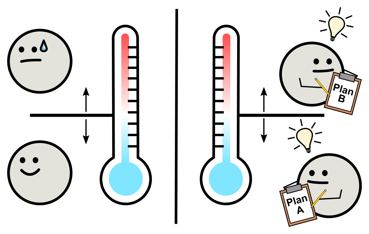
Temperature has a fascinating and complex relationship with biological systems. Intuitively, extreme temperatures can be harmful to living organisms. Anyone who has gotten a sunburn or frostbite before knows what we’re talking about. Because of this, when scientists study how temperature interacts with organisms, they often classify temperature as a potential “stress.”

Yet, temperature is much more than just a potential source of harm. For organisms that lack ears or eyes, temperature is one of the most important sources of information about their surroundings. Temperature is a signal to organisms that their environment is safe or dangerous, allowing them to launch an informed response.

Humans process temperature and respond reflexively, sometimes by shivering in cold weather or by removing a hand from a hot stove. Similarly, a whole range of organisms have intricate temperature-sensing systems. These systems process temperature, along with other environmental signals, to gather information about an environment and help organisms adapt and survive. Thus, temperature plays a crucial role in transmitting life-saving information to biological systems, from helping human cells detect an infection, to aiding bloodthirsty mosquitoes in finding a source of food, to telling microbes when to reproduce, to determining sex in fish.

Temperature Can Signal for Help

When your body temperature reaches just a few degrees above 98.6°F, you may feel like your body is malfunctioning. However, a fever is actually your body’s planned response to a harmful pathogen. A fever signals to organs and tissues that you are fighting an infection, which ultimately leads to responses that help you recover from an illness. Immune fighter cells detect elevated temperature and produce chemicals, some of which directly kill the pathogen and some of which recruit other immune cells.

Activating immune cells in response to a fever helps you recover from an infection, but if your immune cells become activated in the absence of a temperature change, they could cause damage to tissue by killing healthy cells. In fact, unchecked immune cell activation leads to many autoimmune disorders like Type I diabetes and multiple sclerosis. Consequently, temperature is a critical signal for our bodies, but it must be interpreted correctly to truly help us survive.

Temperature Can Signal the Presence of Food

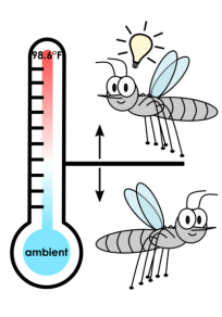
Did you know only female mosquitoes bite? That’s because they need nutrients found in your blood to develop their eggs. In fact, male mosquitoes generally avoid humans. But how do female mosquitoes know where to find us?

Mosquitoes are attracted, first and foremost, to carbon dioxide, which humans constantly exhale. Female mosquitoes also detect movement, odor, and, importantly, heat to find their lunchtime snack. In fact, mosquitoes detect temperature differences as small as a few degrees and avoid objects that are hotter or colder than normal human body temperature. In this case, temperature is among the signals of human blood meal that female mosquitoes detect to navigate to their food.

Temperature Can Signal When Cells Should Divide

The dream of a unicellular yeast is to become two yeast cells. Thus, yeast cells will divide and reproduce until they run out of food or room. However, when yeast cells experience temperatures that they find uncomfortable, they divide much more slowly. If yeast have a single-minded focus on cell division, why would cell division ever slow down?

Some scientists have speculated that high temperatures stress and damage their internal machinery. However, based on recent research in our lab, we’re starting to realize that yeast machinery is not breaking down. In fact, the slower cell division might be not be an unintended consequence, but rather, a planned response.

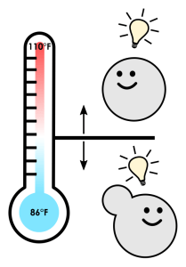
But a planned response to what, exactly? To understand that question, we need to take a bird’s-eye view of yeast, literally. Yeast will grow happily on grapes, where they are fair game for hungry birds. Scientists have shown that yeast can survive being eaten, digested and passed by a bird, which makes birds their primary form of long-distance transportation. Interestingly, a bird’s stomach is the same temperature that causes yeast divide slowly. Putting this together, our lab thinks that warm temperatures might tell yeast that they have been eaten by a bird and that they are currently residing in its stomach. With this information, yeast can shut down their dividing machinery to save energy and resources until they reach a more hospitable environment. Thus, the yeast’s ability to sense temperature and shift into a frugal survival mode is critical for their survival as a species.

Temperature Can Determine Sex

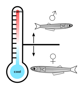
For Atlantic silverside fish, the temperature of a developing egg determines whether the fish becomes male or female. This method of sex determination is drastically different from humans, whose sex is determined by chromosomes. It turns out that this temperature-dependent development is crucial for this species of fish to survive. When female fish are born early in the spring and have time to grow larger, they can hold more eggs. The population of silverside fish therefore benefits from having females born first so they have more time to mature. For this reason, eggs incubated in early-season cool temperatures are predominantly female, while eggs hatched in late-season warm temperatures are predominantly male. (Male fish produce the same amount of sperm regardless of their size, so it doesn’t matter as much when they’re born.) Only 0.04% of eggs become adults, so a small increase in a female fish’s size can have huge consequences for the fish population. In this way, sensitivity to ambient temperature is crucial for Atlantic silverside fish to survive another year.

As we think about the effects of climate change, we should consider animals such as these fish, whose sex ratios vary depending on the seasonal temperature. Rising temperatures could cause fewer silverside females to survive each year, leading to a significant drop in the population. This effect could compound over years, disrupting not only this species of fish but other animals in its ecosystem.

Research Tackles Unanswered Questions About Temperature

A variety of organisms rely on temperature to make decisions about how to best adapt to their environments. However, there are still many unanswered questions about the relationship between life and temperature. For example, how exactly do organisms “sense” temperature on a cellular level? What are their internal thermometers and how do they work? These questions are inspiring for research scientists like us. As we think about the ever-increasing likelihood of a warmer world, we are motivated by the fact that solutions often come from unexpected places. We are hopeful as we look back on the resilience of life through evolutionary time and look forward to a future of greater understanding.

We’ve just published a paper on how a protein’s ability to form hydrogels allows cells to sense and respond to stress.

A bit of background

Like us, cells react to stresses like heat, starvation, and poison. All these stresses cause clumps of molecules to form inside cells. This clumping—aggregation—looks like damaged proteins sticking together. But in our last paper, we showed that instead, these clumps look like they’re a cellular response, not just signs of damage. (See our blog post on it.)

We’re going to go back to the Cellular Institute with its manicured grounds and main building. Happy workers (proteins) go about their often inscrutable business.

The Institute is constantly dealing with awful changes in its environment. The mission is to survive, even to keep growing, during those tough times.

The picture that came out of our earlier study suggested that, overall, the clumping in cells was an organized, reversible process, consistent with a coordinated effort to survive tough times.

But what about for specific proteins? How can you tell if clumping is stress-inflicted damage or an adaptive response? A key test is to see what happens when you stop the clumping during stress. Stopping damage during stress should make cells healthier. Stopping an adaptive stress response, on the other hand, would make cells sicker.

Doesn’t sound too complicated

Easy for you to say, headline! In our study, we had to first find a protein that formed clumps during stress, then figure out how to control its clumping without screwing up a bunch of other stuff (which involved quite a bit of figuring out why it formed clumps, physically, and which bits of the molecule were responsible).

Doing all that required working on the protein in isolation in a test tube, so then we had to put the protein back into cells to see if it worked the same way. Finally, we could expose those cells to stress and see how they responded.

What did we do?

We picked out a specific protein that formed clumps during stress: poly(A)-binding protein, which is nicknamed Pab1 in baker’s yeast, our beautiful and hardy model organism. (Relatives of Pab1 are found in essentially every cell that has a nucleus, from plants to parasites to people.) We purified Pab1, put it in a dish, and exposed it to the same intracellular conditions that follow stress and are thought to cause clumping, heat and pH.

Yeast cells like to live in acidic (low-pH) environments, but keep their insides at neutral pH. When cells get stressed, their internal pH drops, becoming more acidic. That, others have argued, might cause clumping during non-heat stresses like starvation.

What did we find?

First, we discovered that Pab1 “clumping” was something rather specific and wonderful: phase separation into hydrogel droplets.

Phase separation is like a flash mob. Under certain conditions, suddenly, from a mixture of two types of molecules, one type surges together, separating from the other (“demixing”) like oil droplets from water. Bunches of recent studies have shown proteins doing this trick, usually under exotic conditions.

Pab1 phase-separates in response to both pH and temperature, right at the stress-associated ranges of each one. It’s one of the coolest results in the paper: Pab1 is an extraordinarily sensitive sensor of stress, particularly temperature. You might be surprised to know that how yeast cells sense temperature has been a bit of mystery…

Back into the cell

We figured out a way to make Pab1 phase separate at lower or higher temperatures. Remember our key test? That let us make cells with proteins that couldn’t get together during stress. And guess what we found?

Cells with Pab1 molecules that didn’t stick together during stress couldn’t grow during stress. This happened during heat shock, and during starvation, too!

Ultimately, it’s about fitness

There’s been a bunch of beautiful work on phase separation, hydrogel formation, and other quinary phenomena. (Quinary, a term introduced by Stuart Edelstein, refers to a step beyond traditional quaternary structures to structures involving undefined numbers of molecules.) But showing that these phenomena are actually good for cells, as opposed to just gorgeous curiosities, has been challenging. Pab1 delivered for us!

Dive in!

This paper was five years from initial discovery to publication. There’s a lot in there (though the reviewers wanted oh so much more!). For experts, our results are full of surprises. First author Josh has blogged about some of the biophysical aspects and co-first author Chris has blogged about the in vivo aspects.

And there’s much more to do. How, exactly, does Pab1’s phase separation help the cell? We share some ideas in the paper. How, exactly, does Pab1 sense temperature and pH? Watch this space as the story continues to, well, come together.

Hi, I’m Chris. I’m a student in the Drummond lab and coauthor on the paperStress-Triggered Phase Separation Is an Adaptive, Evolutionarily Tuned Response.” When presenting this work, I’ve been asked a few questions repeatedly that I will answer in a FAQ section. I’ll also share an anecdote about the challenges of delineating Pab1’s role in the stress response from its many other functions. I hope the story emphasizes the importance of endogenous protein expression levels.

FAQs:

The ΔP yeast strain grows better at high temperature than the other demixing-impaired mutants – what gives?

How can a few point mutations in the P-domain have a greater fitness effect than deleting the domain wholesale? In figure 6F, when we compare the growth at 40° C of yeast expressing Pab1 variants; mutants “MV->A” and “MVFY->AGPNQ” show a major growth defect. These mutants show a greater defect than the strain expressing Pab1ΔP. This is result is most easily rationalized through the lens of demixing: Pab1ΔP demixes more robustly than either of the mutants; therefore it grows more robustly at high temperature. Figure 6G illustrates the relation between growth and Pab1 demixing. The related question, “how can a few point mutations have a greater effect on demixing than deleting the domain wholesale?” remains open. Josh discusses this point in greater detail in his blog post.

How do mutations to the P-domain affect stress granule formation?

I don’t know. The stress used in figure 6D to trigger Pab1 demixing is too mild to elicit Pab1-marked stress granules in wild type cells (figure 1A). Moreover, while stress granules are easily observed, it is not clear that they are the physiologically functional species. Here, we make the case that perturbing demixing of Pab1 is sufficient to affect physiology. In fact, I favor the hypothesis that quinary assemblies and stress granules are related, but separable phenomena. Pab1 forms sedimentable quinary assemblies in the absence of visible focus formation. Conversely, HSP104 is an example of a protein that colocalizes with stress granules, but isn’t sedimentable.

Is the assembly of other proteins perturbed?

Yef3 is another protein that forms sedimentable species during mild stress conditions. Figure S5 indicates that demixing of Yef3 does not vary between P-domain mutants. However, it is possible that demixing of one of the other ~150 heat-assembling proteins is affected. At its core, this question is about the mechanism of formation of large cellular assemblies. Are all proteins demixing autonomously, or can one protein recruit another to these structures? Is RNA binding sufficient to recruit some proteins? Is recruitment stress specific?

What is going on with RNA?

Right? In vitro we show the following: RNA is not necessary for Pab1 demixing (fig 2E), RNA actually inhibits demixing (fig 2C); and short poly(A) sequences are released during demixing (fig 2A). One interpretation is that demixing is dominated by protein-protein interactions and these interactions are mutually exclusive with RNA binding. In vivo we show that heat-triggered sedimentation of Pab1 is RNase1 insensitive (fig 1B). This result is consistent with the in vitro results, though other interpretations are possible. This model predicts that demixing of Pab1 and poly(A)-positive RNA are not mechanistically connected. I find this unintuitive, but testable.

You speculate about Pab1 as a translational regulator of the stress response. How much is speculation?

If you’re curious about the mechanism by which Pab1 demixing affects stress tolerance, then come to my talk at ASBMBTranslation of heat shock proteins is regulated by poly(A)-binding protein assembly.” Its on Sunday April 23 2017 during the Protein folding, Aggregation, and Chaperones session; or come see the poster later.

Make happy yeast:

Pab1 is an essential gene with a role in many cellular processes. If one wishes to make mutations, delete then replace isn’t an option. Other work has successfully expressed Pab1 from a plasmid. However, cells are very sensitive to Pab1 expression levels – consistent with its known autoregulatory expression mechanism. My first attempt to modify the Pab1 P-domain was with a construct that replaced the native genomic 3’ UTR with a KanMX selectable marker. I also made the control strain with the same marker, but preserved P-domain. The control strain grew slowly and at 30°C and expressed Pab1 at ~20% the wild type. Reduced expression is problematic because demxing is concentration dependent; similarly, over-expression can lead to demixing artifacts. Worse, the proteome of this control strain resembled that of stressed wild type cells, including high chaperone expression. Ultimately, I developed the serial transformation strategy described in the methods (and another blog post). The resulting strains have no selectable markers, native 3’ UTRs, and no extra nucleotide scars. This manor of strain construction solved the reduced Pab1 expression and slow growth problems. Now, I am able to discern the relation of assembly and the stress response, without conflation of growth rate effects.

Hi, I am Joshua Riback, a student in the Biophysical Sciences graduate program co-advised in both the Drummond and Sosnick Labs.

Recently we have published a paper characterizing an intersection between polymer (bio)physics and cell biology. Polymer biophysics elucidates the principles governing the organized demixing of molecules inside of cells. Protein and RNA can demix into massive assemblies such as gels, liquids, and fibers. They lack a fixed stoichiometry so we refer to them as quinary assemblies. We focus on the properties of assemblies composed of poly(A)-binding protein (Pab1) that form in eukaryotic cells in response to cellular stress. We find that the demixing of Pab1 has been tuned for rapid response to stress and is beneficial for surviving that stress. For a little more background, see Allan’s post on our paper.

For those who wish to dive into the paper, please proceed here. Since our story differs significantly from the current results in the field, I have focused this blog post on the polymer biophysics I expected would govern Pab1 demixing vs. reality.

Polymer biophysics of Pab1: Expectations

Prior to obtaining data and based on my reading of the literature in the field, the principles I expected were going to govern Pab1 demixing were:

  1. Pab1 would have an intrinsically disordered region (‘IDR’ or non-foldable low complexity regions) that would allow different Pab1 molecules to flexibly interact in the quinary assembly.

  2. Demixing would be favorable due to multivalent interactions between RNA and Pab1 and between IDRs on different Pab1 molecules.

  3. Pab1’s IDR would promote demixing through interactions between charge, aromatic, or polar residues (i.e. coulomb interactions or hydrogen bonds) and not from aliphatic residues because they are depleted in IDRs. The interacting residues would be enriched in the composition of the IDR.

  4. Pab1’s IDR would be collapsed. When demixed, Pab1’s IDR would interact with other Pab1 molecules; but when monomeric, Pab1’s IDR would self-interact causing it to collapse.

Polymer biophysics of Pab1: Reality

Expectation: #1

Pab1 would have an IDR that would allow different Pab1 molecules to flexibly interact in the quinary assembly.

  • Pab1 has an IDR called the proline-rich domain or P domain.
  • Deletion of the P domain perturbs stress-triggered demixing of Pab1 into quinary assemblies in vitro and in vivo.

Expectation: #2

Demixing would require multivalent interactions between RNA and Pab1 and/or between IDRs on different Pab1 molecules.

  • In vitro, Pab1 demixing does not require RNA; RNA destabilizes demixing. How does this work in cells? Chris considers this further in his FAQ section

  • Although the P domain is the strongest single domain enhancer of demixing, it is not required for demixing. Multivalent interactions between Pab1’s folded domains are required for demixing and govern the salt and pH sensitivity of demixing. When splitting the protein in half, only the half without the IDR can demix.

Expectation: #3

Pab1’s IDR would promote demixing through interactions between charge, aromatic, or polar residues (ie coulomb interactions or hydrogen bonds) and not from aliphatic residues because they are depleted in IDRs. The interacting residues would be enriched in the composition of the IDR.

IDRs are understood by their compositional properties. Based on known IDRs that can demix, important compositional features have been identified (see the review). One of these IDRs, the RGG domain is necessary for demixing of the P-granule protein, LAF-1. The composition of the P domain is different from that of the RGG domain and these other known IDRs implicated in demixing.

For clarity, the compositional plots are fraction residues of each type, and for the evolutionary composition plot, each row is a different species. The hydrophobicity of the residue groups are roughly aromatic, aliphatic, small, polar, and ionizable from most to least hydrophobic.

  • The P domain is strongly depleted for ionizable residues. This composition feature is conserved.

  • The P domain does not have unusual polar or aromatic content. This is conserved.

  • The P domain is unusually hydrophobic; these hydrophobic residues are interacting and stabilize demixing.

Expectation: #4

Pab1’s IDR would be collapsed. When demixed, Pab1’s IDR would interact with other Pab1 molecules, but when monomeric, Pab1’s IDR would self-interact causing it to collapse.

This is a concept drawn from polymer physics 101. If IDR-IDR interactions are sufficiently strong, then the IDR will either collapse if at low concentration or demix if at sufficiently high concentration.

  • The P domain is collapsed. It behaves as expected for a weakly packed, dynamic globule. Extreme mutations in the P domain, such as changing the 15% of the sequence that is glycine to proline or randomly changing the order of ALL the residues in the domain, do not perturb P domain collapse.
  • The extent of P domain collapse correlates with the enhancement of Pab1 demixing in vivo, in vitro, and survival during stress.

Expectation vs. Reality Summary

# Expectation Reality Expectation~Reality
1 Pab1 has IDR IDR (P domain) is conserved in eukaryotes Yes
2 Demixing requires RNA:Pab1 interactions and IDR:IDR interactions Demixing requires only folded domains No
3 IDR promotes demixing with charge, aromatic, or polar residues Net hydrophobicity of amino acid residues enhances demixing No
4 IDR is collapsed. Extent of IDR collapse would correlate with enhancing demixing Size of IDR correlates with the temperature of demixing in vivo and in vitro Yes

Feel free to contact me with questions or comments.

Note: Edward sang the ditty below to us at his going-away party. He was the group’s first postdoc, a scientist of tremendous range who trained as a mathematician and mastered computational molecular evolution, then experimental biochemistry, during his tenure. Now he’s off to start his Marie Curie fellowship in Edinburgh. He is greatly admired, and already deeply missed. – Allan

O Drummond Lab

(to the tune of “Shenandoah”)

O Drummond lab, how I shall miss you,
At the bench and the computer,
And all the fine science that you do.
Away, I must away, across the vast Atlantic.

For five long years I’ve toiled away,
At the bench and the computer,
Measuring proteins and RNA.
Away, I must away, across the vast Atlantic.

All you have taught me so, so much,
At the bench and the computer,
Of biochemistry, evolution, cell biology, statistics, biophysics*, and such.
Away, I must away, across the vast Atlantic.

O Drummond lab, you lovely colleagues,
At the bench and the computer,
And a quintillion heat-shocked yeasties.
Away, I must away, across the vast Atlantic.

*Creative interpretation is needed to fit all these fields in a single line, or single postdoc.

Edward Wallace, December 2015.

We’ve just published a paper on how cells respond to heat stress.

What did we do?

We measured heat-induced protein aggregation and disaggregation in budding yeast, a beautiful and hardy free-living unicellular organism that grows happily at 30°C, gets stressed out starting around 37°C, and dies pretty fast at 50°C.

What did we find?

We discovered that aggregates of endogenous proteins that form after heat stress are fully reversible, form rapidly at specific locations, and in certain cases retain function and fidelity in vitro.

The behavior of endogenous, mature proteins in vivo does not match expectations from studies that use exogenous/reporter proteins or that focus on newly synthesized proteins. In those studies, aggregated proteins are often (but not always) degraded and generally lose their function.

Ideas

What looked like heat-induced protein damage followed by triage and degradation instead looks like an orderly, evolved regulatory response.

Irresponsible speculations

Heat-induced protein aggregation has been seen as mostly physics, but (for proteins that actually aggregate during heat shock) it’ll turn out to be mostly biology. The vast majority of aggregation in the cell in response to heat shock will prove to be beneficial. And not just by preventing toxicity, the way amyloid fibrils are often argued to be beneficial, but by carrying out critical cellular remodeling functions.

Primary among those functions: focusing cellular protein synthesis on mRNAs encoding stress-responsive proteins.

Picture, please?

For the science, there’s no substitute for reading the paper. Here, I want to talk a little bit about how to think about what’s going on in the cell, given our study and everything else we know.

Here is the idyllic campus of the Cellular Institute with its manicured grounds and main building. Happy workers (proteins) go about their often inscrutable business.

institute

When suddenly: catastrophe. A fire breaks out. (Heat shock!) What happens?

What has long been observed—blurrily, as if from orbit, because our instruments aren’t so great—is that suddenly the workers move to particular places and gather together in big groups.

fire-drill-blurry

Then after a little while, emergency crews show up, and some time after that, everything’s back to normal.

So what’s going on?

Most previous work has guessed, for many good empirical reasons, that mainly what’s going on is that the workers are distressed, damaged, even dying. Workers clump together because—gruesomely—heat partially melts them and makes them sticky. Of course, these workers stop working. Some injured workers can be rescued; many of them are beyond saving. The emergency crews triage victims, rescue some, and dispose of the bodies of the rest.

heat-shock-damage

The alternative is that the institute has in place disaster-preparedness plans. What we observe is those plans being carried out to the letter, and the plans for heat shock look like the plans for a wide range of other emergencies:

emergency-plan

Some workers stop working, but not because they’re damaged, just to clear the way for the emergency crew. Others continue to go about their business. Those clumps are groups of workers calmly holding on to each other and collecting at designated assembly points. The emergency crew shows up and tends to the few workers who are actually injured. When they’re done, the crew helps the assembled workers let go of each other and return to their original jobs.

heat-shock-assembly

Our study provides evidence that this latter set of activities describes the behavior of most endogenous, mature proteins. Which is not to say that the damage-and-chaos stuff doesn’t happen! Rather, it happens mainly to newly synthesized proteins and to foreign, destabilized reporter proteins.

So here’s what we think is going on:

fire-drill

The “emergency crew” here is, naturally, the team of heat-induced molecular chaperones.

There’s much more to do, and we’re incredibly excited to share the stories coming out of this work. Stay tuned…

28 May 2015 by Allan

We do plenty of math, so I’d like to test out MathJax support.

Here is an example of MathJax inline rendering — \( 1/x^{2} \). And here is a block rendering: \[ r_{XY} = \frac{\mathrm{cov}(X,Y)}{\sqrt{\mathrm{var}(X)\mathrm{var}(Y)}} \]

Now, if we’d like to get serious, we’d do something involving multiline aligned equations, like \[ \begin{align} \mathcal{N}(t, \mu, \sigma) &= \mathrm{normal} \newline &= \frac{1}{\sqrt{2 \pi} \sigma} e^{-\frac{(t-\mu)^2}{2 \sigma^2}} \end{align} \] or even an inline formula like \( \sum_{t=0}^{\infty} \frac{x^t}{t!} = e^x\).

Or we could try defining a command, like this. \( \newcommand{\water}{\mathrm{H}_{2}\mathrm{O}} \)

Buffer slides off the sides of our tubes like \(\water\) off a duck’s back.

22 Apr 2015 by Allan

Our new lab website is up and running. My goal with this site is to change how we communicate our science to the world, and to help others to get over barriers to that change.

In terms of technology adoption, I believe we are early majority types. That means the technology we’re using is relatively mature–we’re not risking much–but most people in our sphere aren’t yet using it. If you’re looking for innovation or are an early adopter, you’ll find this site to be hopelessly outdated!

Open

The site is fully open source, available in a GitHub repository.

Connected

Just as critical scientific interactions have taken place by correspondence and at scientific meetings, many such interactions now take place online in social media. Data and figures now have homes in online repositories. (This stuff is forehead-slappingly obvious to some, and news to others.)